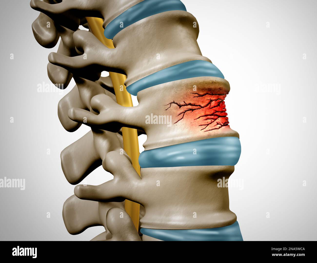
Lumbar Compression Fracture, Illustration - Album alb3774451
By A Mystery Man Writer
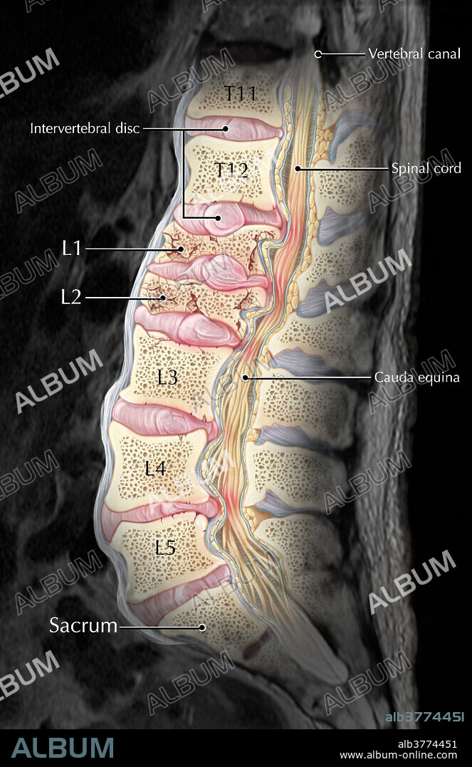
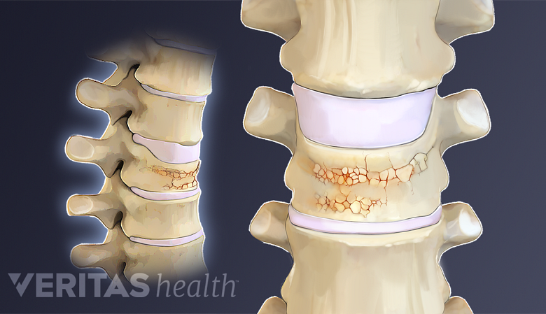
Download this stock image (alb3774451) from - An interpretive illustration of an MRI depicting a sagittal view of compression fractures at the L1 and L2 vertebrae as a result of osteoporosis. Over time as bone becomes weaker and more porous, they become more susceptible to injury and fractures, especially in situations where significant weight or stress is placed on the bone. In this case, the vertebral bodies of L1 and L2 have collapsed, resulting in a displacement of the bones and intervertebral discs into the spinal canal, resulting in pain and possibly reducing the patient's mobility.

COMPRESSION - Stock Photos, Illustrations and Images - Album

Lumbar spine fracture, Radiology Reference Article

Compression fracture of the fourth lumbar vertebra of a calf

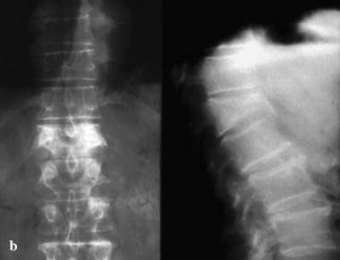
Lumbar spine compression fracture, Radiology Case

Lumbar Compression Fracture, Illustration - Stock Image - C027

Spinal compression fracture Radiology Reference Article
Compression fracture spine hi-res stock photography and images - Alamy

2,934 Compression Fracture Royalty-Free Photos and Stock Images

compression fracture lumbar L4-5, S1 level with loss space of disc

Lumbar Compression Fracture, Illustration - Album alb3774451

COMPRESSION - Stock Photos, Illustrations and Images - Album
Simple Compression Fracture (Case 16) - Clinical Imaging of Spinal
Lumbar Compression Fracture - Summit Spine

2,934 Compression Fracture Royalty-Free Photos and Stock Images

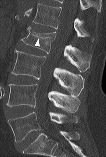
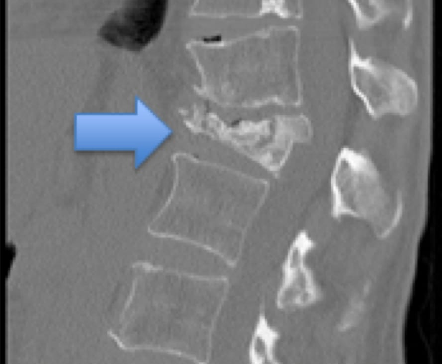
CT Scan of Lumbar Vertebral Column With L1 Vertebral Compression

- for UNISTAR China ZBD-04A Tank Digital Camo RANDOM NUMBERS 1/72

- xiaxaixu Men's Tear Away Pants, Loose Fit Basketball Pants High Split Snap Button Sweatpants

- Lucy Capri

- hometrends 400 Thread Count 100% Cotton Sheet Set - Sateen, Size: Twin - King

- Baretraps Salina Tall Wide Width Wide Calf Knee High Boot