Optical Coherence Tomography: Imaging Mouse Retinal Ganglion Cells In Vivo

By A Mystery Man Writer
Scientific Article | Structural changes in the retina are common manifestations of ophthalmic diseases.

OCT imaging leaps to the next generation

Fig. 9.10, [In vivo confocal reflectance and]. - High Resolution Imaging in Microscopy and Ophthalmology - NCBI Bookshelf
In vivo volumetric imaging of human retinal circulation with phase-variance optical coherence tomography

Optical Coherence Tomography: Imaging Mouse Retinal Ganglion Cells In Vivo
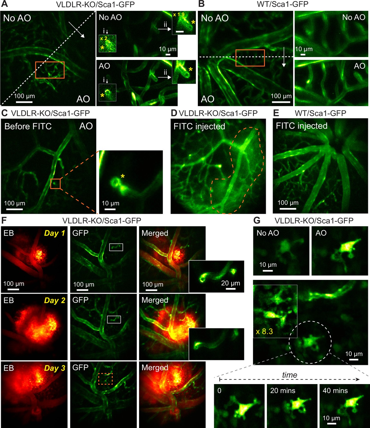
Retinal microvascular and neuronal pathologies probed in vivo by adaptive optical two-photon fluorescence microscopy

Imaging - Experimental Glaucoma & Imaging Laboratory - Dalhousie University

Mouse Retina OCT Fibergram Alignment with Confocal Images

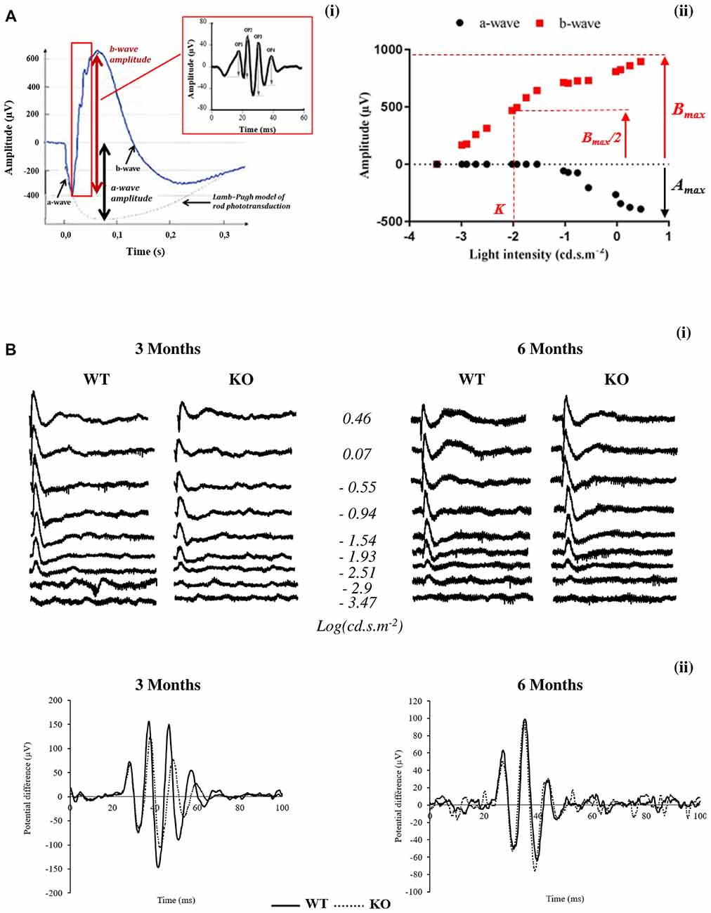
Topical Nerve Growth Factor (NGF) restores electrophysiological alterations in the Ins2Akita mouse model of diabetic retinopathy - ScienceDirect
Optical Coherence Tomography: Basic Concepts and Applications in

Optical Coherence Tomography: Imaging Mouse Retinal Ganglion Cells In Vivo

Imaging - Experimental Glaucoma & Imaging Laboratory - Dalhousie University

Genes, Free Full-Text
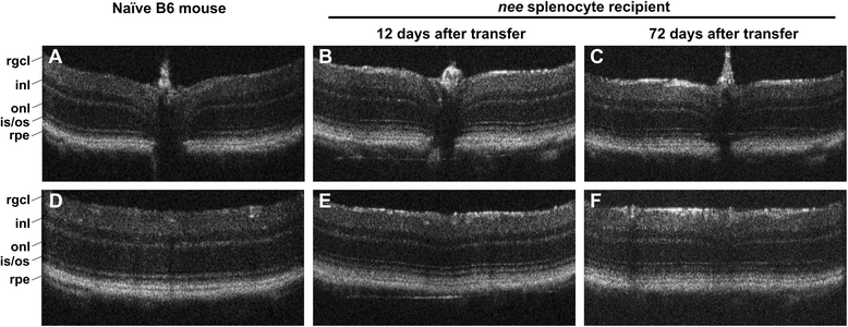
Adoptive transfer of immune cells from glaucomatous mice provokes retinal ganglion cell loss in recipients, Acta Neuropathologica Communications
Frontiers Early Retinal Defects in Fmr1−/y Mice: Toward a Critical Role of Visual Dys-Sensitivity in the Fragile X Syndrome Phenotype?
- Induzido Para Motores De Avanço Align

- TBMáquinas - Turcite para Barramento - São Caetano do Sul, SP - Blog - Induzidos para Avanço Automático
- ALINHADOR DE RODAS CELFER-27040 (12328)

- Calços Para Cambagem em Y D10/D20 Medio 50 peças - Artnel

- Relator quer arquivar processo contra deputado que espalhou fake news sobre Marielle


/data/mothercare/24dec2021/NB372-2.jpg)